Exploring Science, Technology and Art
#ERN2023 Photo Competition Gallery
The #ERN2023PhotoCompetition was part of the European Researchers’ Night 2023 initiative. This was an open competition and everyone was encouraged to submit up to two original photographs in the following category:
→ ‘Science for Everyone – Sustainability and Inclusion’ (nano/micro images obtained through microscopes, images related to the labs, setups, sample preparation, simulations, everyday life related to science, etc.)
Your talent and unique visualisations were honoured and celebrated.
→ Gallery 2023
Hélder FONSECA
PHOTO 30 2073: The ghost yellow room
This picture was taken inside the INL cleanroom, in the photolithography bay. This room is yellow because some used chemicals are not sensitive to their wavelength, so it prevents unwanted resist exposure and development.
This picture is a long composite of stacked short exposures, done with just 2 persons wandering across the area. This creates a sense of blur motion and a ghosting effect.
FREDILSON MELO
PHOTO 52 Bloom, Algae! Bloom!
Algal bloom in a tray used for growing rice for research.
This photo was taken inside a walk-in growth chamber at ITQB NOVA during rice cultivation. Due to the soil's nutrient richness and good quality of light, the algae multiply, covering the whole tray. This is not problematic since rice can still grow normally in a pot that separates it from the algae. The purple-ish colour of the photo is due to the light spectrum used to grow rice.
Tânia Marques
PHOTO 21 Mold on Pear I
The photo was taken at home and is a macro shot of mold on a pear.
TIAGO OLIVEIRA
PHOTO 13 Bridge
SEM picture of a suspended membrane on an etched silicon wafer, from a destroyed MEMS device.
In an ode to the value of destruction, this device, in order to be fully inspected, has to have its layers selectively removed so that they can all be inspected, reaching the end, where only the bottom membrane stands. As if to invoke the Moria bridge, where Gandalf fought the Balrog, the rough walls of the silicon indicate a deep fall into the void.
DIANA alves
PHOTO 17 Twinkle twinkle little star, nano-wonders made close from afar
This is an SEM picture of zinc oxide synthesised under the scope of my research project with a magnified star-like structure. As we do with the microscope, we should be able to zoom in on the problems of this world so they can turn into wonderful opportunities for us all.
The picture was taken at INL, using Quanta FEG 250 Field Emission SEM, magnification: 5000X.
Maciej Majdecki
PHOTO 46 Solid versus solution under UV light
The photograph shows an organic compound in the form of a solid and a solution that has been exposed to UV radiation. We can see here a variety of colors depending on the state of matter.
The photo was taken in the laboratory of the Institute of Chemistry of the Polish Academy of Sciences in Warsaw.
MAFALDA NETO
PHOTO 47 Mötley (Epithelial) Crüe
Intestinal organoid-derived monolayer observed under confocal microscope (20X magnification). Nuclei in blue, cytoskeleton in red, cell tight-junctions in green.
JOÃO AZEVEDO
PHOTO 28 "From Simple Shapes, Science Awakes"
This photo is a TEM image of a Magnesium salt structure on a sample solution that was created upon my Master's Thesis work on DNA origami. I colored the image to enhance the specific planar features of the cubic salt crystals, separating them from the aqueous background.
Maciej Trzaskowski
PHOTO 2 "The secret garden"
The image shows Rhododendron pollen grains exhibiting pollen tubes. The pollen tube is a pipe that transports the male gamete cells from the pollen grain in the process of plant reproduction.
The image was taken with the use of a Hitachi SU8230 scanning electron microscope owned by the Centre for Advanced Materials and Technologies CEZAMAT, Warsaw University of Technology, magnification: 3000X.
Maciej Trzaskowski
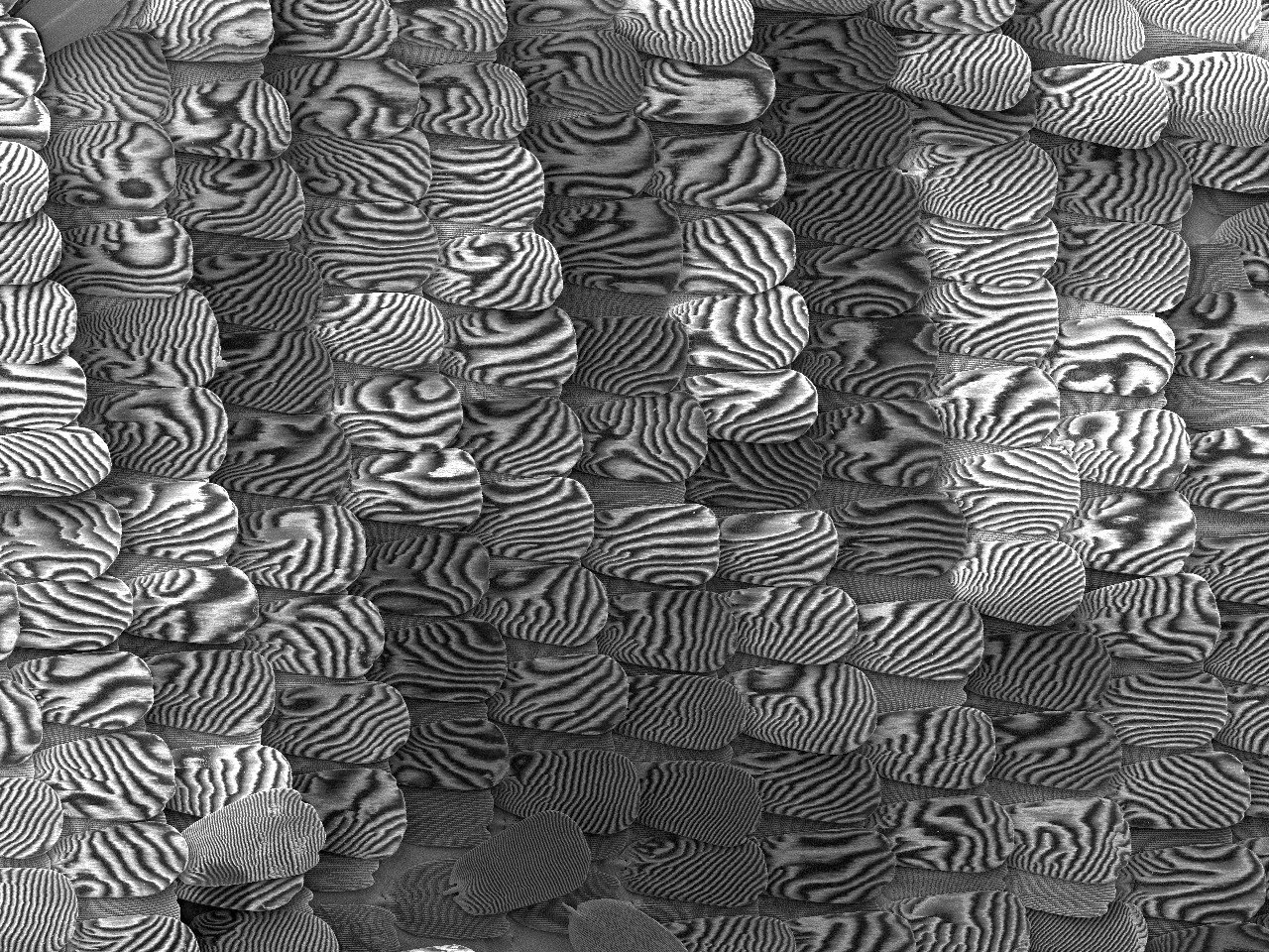
PHOTO 1 "Effect of the Scales"
The image depicts scales found on the wings of the Purple Emperor (Apatura iris) butterfly. These scales showcase a grid-like microstructure with varying grid spacing between layers. As a result, an iridescent effect emerges, causing colours to gradually change as the viewing angle shifts while observing the butterfly's wing on a macro scale. Under specific conditions, as illustrated in the image, the grid gaps overlap, giving rise to the Moiré effect – interesting interference patterns.
The image was taken with the use of a Hitachi SU8230 scanning electron microscope owned by the Centre for Advanced Materials and Technologies CEZAMAT, Warsaw University of Technology, magnification: 120X.
CARLA GASPAR
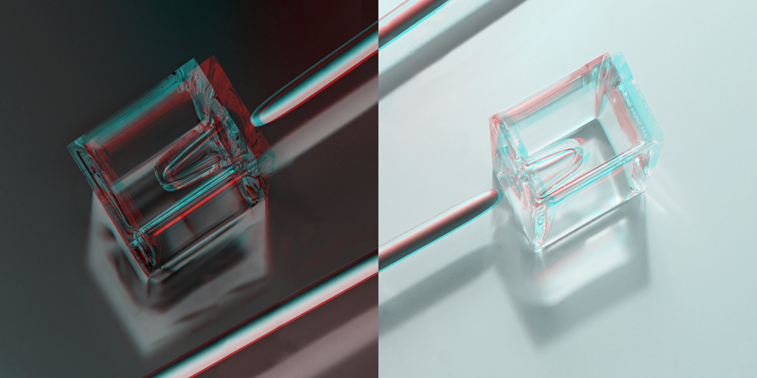
PHOTO 3 Iminente#5
Iminente #5 is part of a series made up of a set of objects, photographed in a stereoscopic phantogram, in anaglyph, to be seen (with red and cyan filter glasses) at 45 degrees, horizontally, so that the projected figure, see, in 3D, vertically, suspended in the air. The objects represent the state of suspension, stopped in time and space, contradicting the gravity of their weight. The piece enables interaction and duality between the two bodies – the Virtual and the Physical.
Fragility and Resistance. The series does not have a limit on the number of objects. It is a work that will be continued over time.
The photo was taken in my studio.
JOSÉ FERNANDES
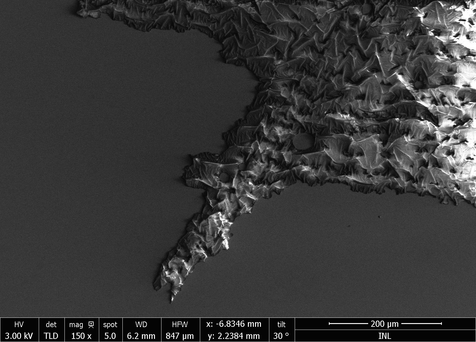
PHOTO 4 Spiky Silicon Donut
Polymerized photoresist after being exposed to high-density plasma. The hardness of the material makes it difficult to etch after a long process.
Since it is a non-conductive material in an SEM system, creates this light and shadow effect.
This photo was taken in a NovaNanoSEM 650 inside the INL Cleanroom.
JOSÉ FERNANDES
PHOTO 5 Rocky "Polymer" Mountains by the Sea
Patterned Tall Silicon Grass with 50 micrometers.
Silicon Grass or Black Silicon is a common microfabrication technique with several advantages, like an increase in the efficiency of a photovoltaic solar cell.
This photo was taken in a NovaNanoSEM 650 inside the INL Cleanroom.
GEORGE JUNIOR
PHOTO 6 Organic channels through to highland tubes
Carbon Nanotubes by Scanning Electron Microscope using a CVD with hot-wall approach.
XAVIER PINHEIRO
PHOTO 7 A glimpse of the unknown
The optical microscope image shows the edge of a decorative metal part coated with a PVD multilayer that has been subjected to a harsh environment (Neutral Salt Spray test) for 200 hours.
But what can really be seen there? A future to be discovered in a distant celestial body where mankind can thrive or a post-apocalyptic planet Earth that humanity did not prevent from happening.
XAVIER PINHEIRO
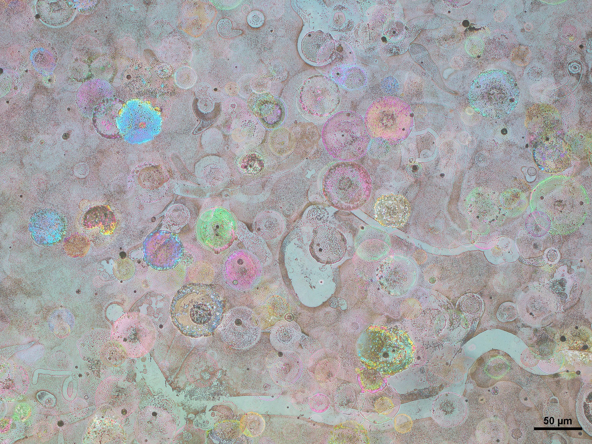
PHOTO 8 Wilson the Substrate
Spincoating deposition of an organic thin film used as a transport layer in solar cells, can be challenging, especially if it is not optimized. However, there are times when an experiment unintentionally creates a company.
XAVIER PINHEIRO
PHOTO 9 Oh! Hi friend!
During the spin-coating deposition of an organic thin film, used as a transport layer in solar cells, some strange marks can appear. Or, for example, we may receive a visit from a friendly bubble that brightens up a hard day's work.
ANA PINHEIRO
PHOTO 11 The Big Bang Theory
This photograph displays a glass sample with a deposition from colloidal perovskite nanocrystals in solution.
The deposition was made in a spin-coater, originating a very interesting centrifugal spread of the luminescent nanocrystals. Under ultraviolet radiation, the radial arrangement of the two different light-emitting species, one blue and one green, creates a composition that is reminiscent of the vast universe from which Earth appears as a very small single dot.
Laboratory of Photochemistry, Department of Chemistry, from FCT/NOVA. February of 2019.
ANA PINHEIRO
PHOTO 12 Where is Earth expanding to?
It is critical that we accelerate our research on uncovering the metamorphosis of matter into energy if we are to re-birth a new, more sustainable, universe.
In this composition, we can see three glass samples with a deposition of a colloidal perovskite nanocrystals thin-film, a promising material from the energy generation field. The lack of homogeneity of the sample and the thermal instability of this system originated in a swirling composition of two different light-emitting species. When under ultraviolet light, we are able to see a compound piece that very much resembles our planet, while being a metaphor for how solar energy can shine a new light upon us, as a consumer’s society.
Laboratory of Photochemistry, Department of Chemistry, from FCT/NOVA. February of 2019.
TIAGO OLIVEIRA
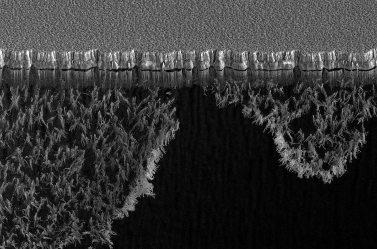
PHOTO 12 Moss
SEM picture of fluor residues from reactive ion etches, suspended in the air after the final release of a device.
As if moss hanging from a tree, this carpet of fluor residues hangs from an aluminium structure, so thin and loose that it ruptures itself with just airflow. An almost warm and lush beauty, contrasting with the cold and barren aluminium structures.
TIAGO OLIVEIRA
PHOTO 14 Out of Focus
An optical microscope view through an etched wafer, focusing on the structures on the other side.
While finishing up the inspection on a final silicon etch, I turned off the microscope light and kept talking to someone. During that time, the microscope software was set to AUTO EXPOSURE. With no additional light sources, the software kept pushing the capture time and eventually settled on this view, which I found quite beautiful and ethereal. Almost alien-like.
Pavlína Vávrová
PHOTO 15 The Mystery of Living Together I
The picture of dual-species biofilm of methicillin-resistant Staphylococcus aureus and Candida albicans cultivated 24 h in Soybean medium supplemented by human plasma. Biofilm was stained by Syto 9, propidium iodide, and calcofluor white.
The picture was taken in the summer of 2023 with an Olympus Provis AX-70 epifluorescence microscope equipped with a camera device (DS-Fi3, Nikon, Japan) and NIS Elements software. Charles University, Faculty Of Pharmacy, Czech Republic
Pavlína Vávrová
PHOTO 16 The Mystery of Living Together II
The picture of dual-species biofilm of methicillin-resistant Staphylococcus aureus and Candida albicans cultivated 24 h in Lubbock chronic wound medium. Biofilm was stained by Syto 9 and calcofluor white.
The picture was taken in the summer of 2023 with an Olympus Provis AX-70 epifluorescence microscope equipped with a camera device (DS-Fi3, Nikon, Japan) and NIS Elements software. Charles University, Faculty Of Pharmacy, Czech Republic
DIANA ALVES
PHOTO 17 Twinkle twinkle little star, nano-wonders made close from afar
This is an SEM picture of zinc oxide synthesised under the scope of my research project with a magnified star-like structure. As we do with the microscope, we should be able to zoom in on the problems of this world so they can turn into wonderful opportunities for us all.
The picture was taken at INL, using Quanta FEG 250 Field Emission SEM, magnification: 5000X.
DIANA ALVES
PHOTO 18 It’s always darkest before the dawn
This is an SEM picture of zinc oxide synthesised under the scope of my research project. As the picture suggests with a transition from a darker to a brighter area, we should be reminded that together it’s easier to find the silver lining of this ever-changing and ruthless world.
The picture was taken at INL, using Quanta FEG 250 Field Emission SEM, magnification: 5000X2020) of the Portugal 2020 Program [Project No. 45073, "ITEC Smart Automation I4.0"; Funding Reference: NORTE-01-0247-FEDER-045073.
ENZO RIBEIRO
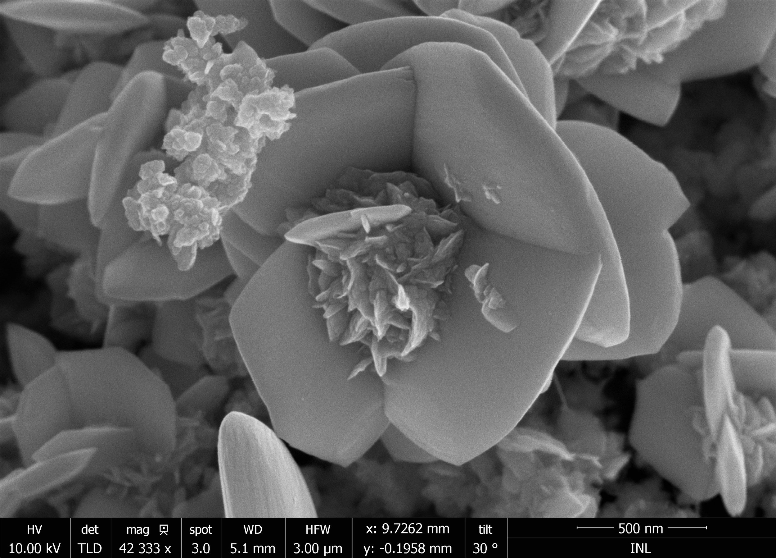
PHOTO 19 A flower made of Bi2Se3 petals
This image was taken at the clean room SEM and it is a crystallized phase of the Bi2Se3 material aimed to be used in solar cells. This material begins to grow as "blades" and repeatedly grows transversely to itself, creating what can be interpreted as petals, ultimately building itself into a flower-like structure.
ENZO RIBEIRO
PHOTO 20 The blooming garden
This image was taken at the clean room SEM and it is a crystallized phase of the Bi2Se3 material aimed to be used in solar cells. This material begins to grow as "blades" and repeatedly grows transversely to itself, creating what can be interpreted as petals and/or wings, ultimately building itself into flower- and butterfly-like structures. Several of these structures can be seen in this image, therefore making this a garden full of flowers and butterflies.
TÂnia Marques
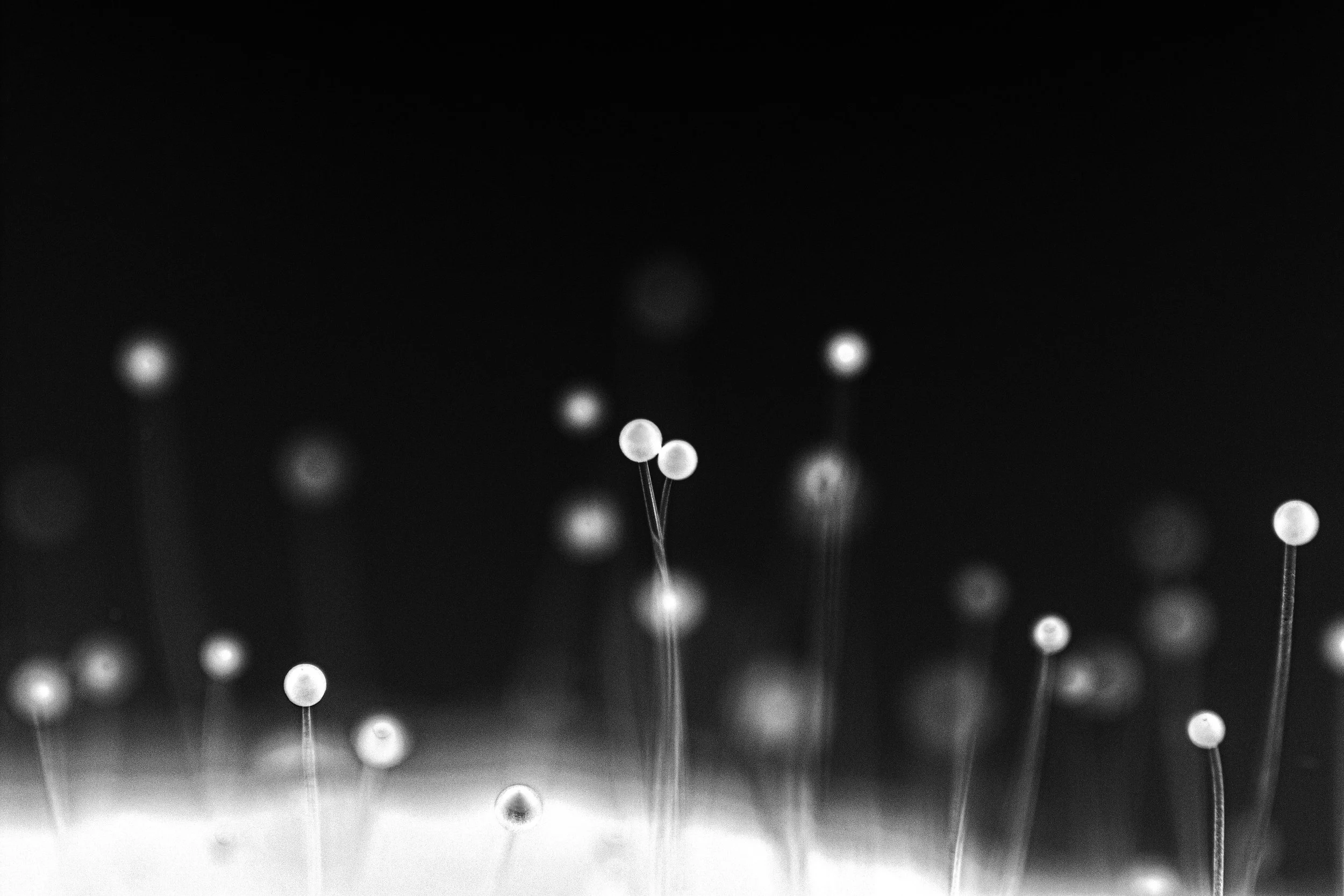
PHOTO 22 Mold on Pear II
The photo was taken at home and is a macro shot of mould on a pear. The photo was taken in colour, transformed to black and white and then to the negative.
VÍTOR LOPES
PHOTO 23 Pollock inspired FTO
The optical microscopy image presents an intricate and dynamic arrangement of deposited FTO layers on the substrate by spray pyrolysis, reminiscent of a Jackson Pollock masterpiece. This description blends the scientific aspects of the microscopy image with the artistic qualities it evokes, drawing parallels to Pollock's style to create a vivid and engaging narrative.
Thin, wispy lines of FTO material crisscross the substrate in a seemingly random but captivating pattern, resembling Pollock's signature 'drip painting' technique.
This image was taken in an OM presented in the TEC group lab.
Gemma Rius
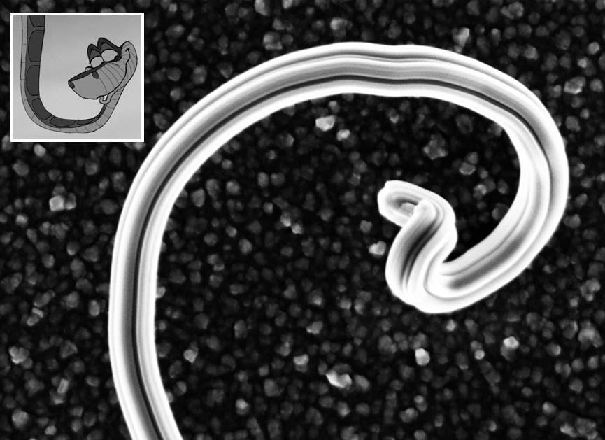
PHOTO 24 The Charm of the Nano-Snake
The photo captures a sample dipped into an electroplating solution to deposit Cu(In, Ga)Se2, a solar cell absorber material. The right-hand side of the sample was dipped into the electrodeposition solution (one world), while the left-hand side is where the metal contacts established an electrical connection (the other world).
I took this photo in the INL cleanroom using an optical microscope. The sample was used to fabricate micro solar cells.
ANDRÉ VIOLAS
PHOTO 25 Tagliatelle alla Vi
This is a scanning electron microscope (SEM) image taken in the middle of a lithography step with a double resist layer to obtain a novel dielectric/metal line architecture to be implemented on the rear contact of bifacial CIGS solar cells.
ANDRÉ VIOLAS
PHOTO 26 Yellow life at the cleanroom
Many of the nanofabrication procedures are performed inside a cleanroom environment due to the very small features and the need for the control of the presence of defects. Specifically in lithography processes, a clean environment is essential, and some of the polymers used are sensitive to ambient light. Some of the developed work occurs with the yellow light shown in this image.
This photo shows the conditions of a typical nanofabrication day.
Olesia Dudik
PHOTO 27 Ocean Coast
The image depicts the edge of a plasma-treated nickel/immersion gold electrode taken through a FEI Quanta 650 FEG scanning electron microscope (AEMIS Facility, INL) at 1780x magnification. The scale bar shows 25 μm.
HÉLDER FONSECA
PHOTO 29 "Baby, strike my fire!"
The moment of plasma striking inside a sputtering system. The purple glow is obtained by the dissociation of argon gas with the help of an RF voltage. This is performed in a chamber pumped down to an ultra-high vacuum.
The photo was taken with a standard camera, through a thick glass window.
ERAY ER
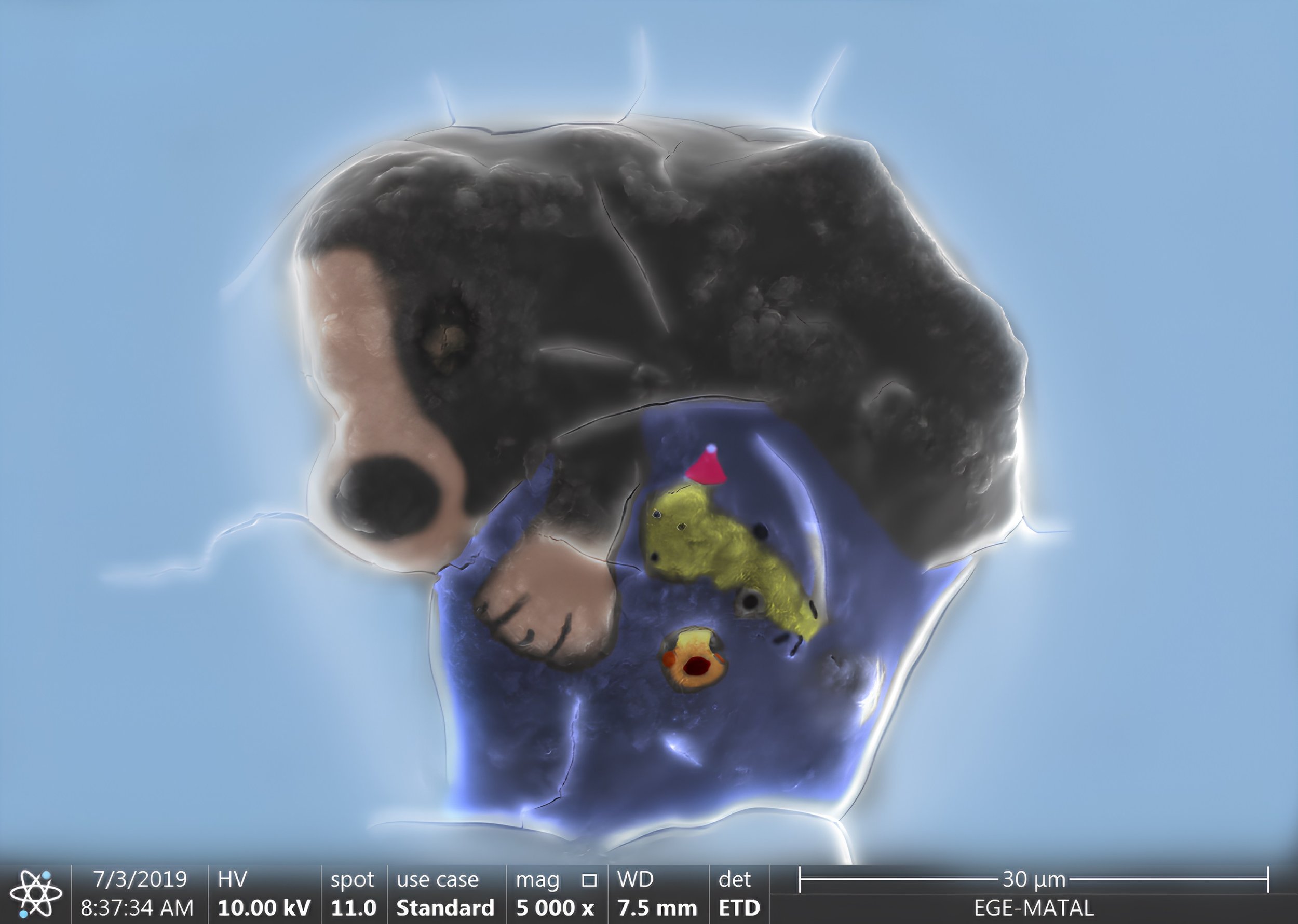
PHOTO 31 Our Loyal Friend
It is a scanning electron microscope(SEM) image obtained after covalent immobilization of the peroxidase enzyme obtained from horseradish on the surface of cellulose acetate nanofibers designed for the determination of hydrogen peroxide from free radicals.
Thermo Scientific Apreo S was used as the SEM device at the Central Research Test and Analysis Laboratory Application and Research Center at Ege University.
Jitendra Kumar
PHOTO 32 Silver Nanodendrites: Nature's Winter Elegance
This captivating scanning electron micrograph unveils the exquisite silver nano dendrites made by electroless chemical deposition on a single-crystal silicon substrate at the Indian Institute of Technology Kanpur, India. Resembling delicate leaves on a winter's day, these nanodendrites branches akin to the elegance of Christmas trees.
Each nano dendrite branch mirrors the symmetrical allure of snowflakes, showcasing the fusion of scientific precision and natural aesthetics. As we explore this miniature forest of nano dendrites, we are reminded of the fascinating world that exists beyond the naked eye, where science and art harmonize to reveal nature's craftsmanship at its finest.
Jitendra Kumar
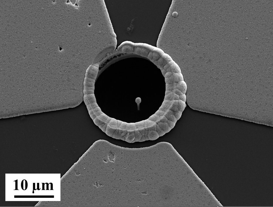
PHOTO 33 Silicon Nanowires with Nickel "Nano Lollipop"
Microfabrication artistry made at the Indian Institute of Science and Indian Institute of Technology Kanpur, India. At its foundation lies a silicon trench insulated by a silicon nitride layer. Nestled atop this insulating silicon nitride layer are three resplendent gold electrodes encircling a silicon nanowire.
Silicon nanowire rises gracefully from this composite base, crowned with a delightful surprise. At its summit, the nanowire has sphere-shaped nickel deposited by electrodeposition, which resembles miniature "nano lollipops."
This silicon wonder, mere nanometers in dimension, is made using deep reactive ion etching. The addition of spherical nickel brings a touch of whimsy to the world of nanotechnology while enhancing the potential for applications ranging from advanced electronics devices to nanosensors.
Buse Naz Gürbüz
PHOTO 34 Crystal Blossom
he image of citrus fibre and water mixture under a microscope 20X (100μm).
This photo was taken in the INL facilities using NBI - Nikon Ni-E Upright Widefield/Fluorescence Microscope in the polarized mode.
JOÃO CUNHA
PHOTO 35 Energy Powder
This Scanning Electron Microscope image shows battery-grade Lithium-Iron-Phosphate particles of sub-micrometer size (about a thousand times smaller than a human hair), randomly arranged within a battery powder. Lithium-iron-phosphate is a cathode material employed in a considerable share of lithium-ion batteries available in the market. The size, shape and microstructural arrangement of the particles within a battery electrode will ultimately influence performance, and as such characterization and control over battery manufacturing, all the way down to electrode microstructure is paramount to improve the performance of lithium-ion batteries. The white bar length corresponds to 1 micrometre.
This micrograph was taken at the INL - International Iberian Nanotechnology Laboratory Micro and Nano Fabrication facility using a FEI NovaNanoSEM 650. This research has received funding from the European Union’s Horizon Europe research and innovation programme under the Marie Skłodowska-Curie grant agreement No. 101062008.
JOÃO CUNHA
PHOTO 36 Microscale River Basins
This Optical Microscope image shows a metallic thin film of Titanium-Tungsten alloy sputtered over a polymer layer. When subjected to moderate temperatures, the Titanium-Tungsten film cracks due to internal mechanical thermal stresses. The cracks propagate along the film forming channels akin to river basin canals, but with micrometer-sized channels. The colours result from the interaction of light with the thin film.
This image was taken at INL - International Iberian Nanotechnology Laboratory Micro and Nano Fabrication facility using a Nikon Eclipse L200N.
PEDRO REBOLA
PHOTO 37 Ancient Alphabet
This is an SEM image of the thermal dewetting of periodic Au nanodiscs on an ITO substrate. The roughness of the substrate and the discontinuity of the nanodiscs caused the Au to diffuse in different directions and form these strange shapes that I am calling an ancient alphabet. Since the nanodiscs were periodically deposited, these structures are also periodic.
This image was taken using the cleanroom SEM at INL - International Iberian Nanotechnology Laboratory.
PEDRO REBOLA
PHOTO 38 The Surface of a Distant Planet
This is a SEM image of a thin Au film that was deposited on top of a resist structure that fell down at periodic spots, because of the weight. Two resists were initially deposited and a nanoimprint lithography process was done to open periodic holes in the top resist. Then the bottom resist was developed through the holes and a thin Au film was deposited. Since there was too much development, the top resistance fell, creating the effect seen in the image. For me, this image really seemed like an image taken off the surface of a distant planet.
This image was taken using the cleanroom SEM at INL - International Iberian Nanotechnology Laboratory.
Andrea Capasso
PHOTO 39 Wee Book
A wee book waiting to be browsed!
Ti3C2Tx MXene nanosheets were obtained by exposing Ti3AlC2 to a strong acid.
Original, untreated SEM image - FEI Quanta 650 FEG, operated by Siva Sankar Nemala.
Andrea Capasso
PHOTO 40 Lil Garland
A little garland for a warm celebration!
ZnO nanorods grown on ITO using hydrothermal synthesis
Original, untreated SEM image - FEI Quanta 650 FEG, operated by Siva Sankar Nemala.
JOÃO AVÓ
PHOTO 41 Shattered Light
Unlocking the Spectrum of Sustainability and Inclusion: Behold the luminescent polymer films, each a beacon of hope on a black canvas of possibilities. These films imbued with TADF emitters, hold the key to sustainable lighting solutions. In this image, science meets art, showcasing how innovation lights the path to inclusion for all.
This picture was taken in my previous lab, at Instituto Superior Técnico, Lisbon.
ELMAHDI AMAR
PHOTO 42 High atomic resolution STM image of Bi2Se3 at room temperature
In this work, an electrochemical etching was optimized to get an adequate tip with a cone shape in the apex. One of the best sharp W-STM tips was efficient in obtaining high-resolution scanning tunnelling microscopy (STM) images. We managed to measure the atomic resolution STM image (5.00 nm ⅹ 5.00 nm) of a Bi2Se3 surface obtained with a home-made STM in air at room temperature (Bias voltage, V=1.6V, Tunneling current, I= 582pA).
The STM image was obtained at the SPM lab at INL - International Iberian Nanotechnology Laboratory.
ELMAHDI AMAR
PHOTO 43 Fourier transform spectra corresponding to the STM image
The FFT of the corresponding STM image seems like goldfish eyes in a dark cave in the ocean. The STM and FFT-filtered images illustrate clearly the hexagonal symmetry of the surface lattice. However, in the STM image, there is a region where we can´t clearly see the structure of the surface because atoms easily jump regularly between the sample and the STM tip. In addition, there is a high diffusion of atoms as the measurements were performed at room temperature.
The FFT image was achieved using WSxM software at INL - International Iberian Nanotechnology Laboratory.
FILIPA COSTA
PHOTO 44 Incubating
African Violet Cultivation.
Photograph taken at the Herbarium of the University of Aveiro.
Maciej Majdecki
PHOTO 45 Frozen flask with fluorescent material
The photo shows a flask with a solution of a fluorescent substance under UV light that has been cooled to a temperature below -70 Celsius degrees to start the crystallization process.
The photo was taken in the laboratory of the Institute of Organic Chemistry of the Polish Academy of Sciences in Warsaw.
Claudia Ruiz
PHOTO 48 Inner light
I like macro nature photography because of the fascination and amazement that it generates both in me and in the viewer to discover worlds that the eye does not capture with the naked eye.
Seeing a Hypericum flower under a UV light and discovering the circulatory system makes me fall even more in love with the wonderful natural world around us and intensifies the desire to share these natural wonders.
Claudia Ruiz
PHOTO 49 Under the light
I like macro nature photography because of the fascination and amazement that it generates both in me and in the viewer to discover worlds that the eye does not capture with the naked eye.
Seeing small Helichrysum bracteatum seeds under UV light and discovering that luminescence makes me fall even more in love with the wonderful natural world around us and intensifies the desire to share these natural wonders.
RAFAEL PINTO
PHOTO 50 On the tiny scale of a butterfly
A closer look at a butterfly’s wings reveals an intricate and colourful coat of tile-like scales, layered side-by-side creating some of the most beautiful patterns in the animal world. Besides their astonishing looks, these insects play an essential role in our ecosystems and provide us with many natural resources such as food, by aiding plant reproduction through pollination. If we desire a more sustainable future, it must include these amazing creatures.
Our lives depend on the role of pollinators such as butterflies, and it is vital to stop their decline to have prosperity in the upcoming times.
The photo shows a close-up view of the hindwings' scales of the swallowtail butterfly Papilio machaon and it was taken using a digital microscope.
Fredilson Melo
PHOTO 51 The Red Plant
Red-fluorescent protein (RFP) on the membrane of cells in the leaf of a Nicotiana benthamiana plant.
This photo was taken on a fluorescence microscope at ITQB NOVA during an assay to understand where a protein (tagged with RFP) is located within the cell. Determining the location of this protein is important to understand how the protein works during plant response to stressful conditions.
Neha Kumari
PHOTO 53 Microscopic Beauty: The Silicon Nanowire Flower
This backscattered electron image reveals an attractive silicon nanowire structure resembling a delicate flower. The image was taken at the Indian Institute of Technology Kanpur (IITK), India.
This flower emerged as a result of metal-assisted chemical etching of a silicon wafer. The central circular area represents porous silicon, while the surrounding regions resemble dandelion flowers, showcasing the silicon nanowires.
The bright white particles scattered across the surface region of these nanowires are silver particles.
Ihsan Çaha
PHOTO 54 Atomic resolution HAADF image of FeCo2O4 nanoparticle
High-angle annular dark-field scanning transmission electron microscopy (HAADF-STEM) image of FeCo2O4 nanoparticle. This image was acquired from a probe-corrected FEI Titan G2 Cubed ChemiSTEM, operating at 200 kV Laboratory.
Fresh samples for STEM analysis were prepared by dispersing the powder mixture in ethanol and drop-casting it onto a holey carbon Cu grid.
JULIANA SOUSA
PHOTO 55 The perfect world of the cookies monster
This photo is a SEM image of metal organic compound. This nanomaterial was employed in adsorbing organic pollutants found in wastewater.


